Macular hole is a condition that affects the macula, the central part of the retina responsible for our sharpest, most detailed vision. It occurs when a small break or opening develops in the macula, which can cause distortion or loss of vision in the center of the visual field. Macular hole usually affects people over the age of 60, and is more common in women than men.
There are four stages of macular hole, which are classified based on the size and severity of the hole, as well as the degree of damage to the surrounding retina.
Stage 1: Foveal detachment
The first stage of macular hole is foveal detachment, which occurs when the macula starts to pull away from the underlying retinal tissue. At this stage, the hole is very small and may not cause any noticeable symptoms. However, if left untreated, the hole can progress to the next stage.
Stage 2: Partial-thickness hole
The second stage of macular hole is a partial-thickness hole, where the hole extends through the outermost layer of the retina but does not reach the underlying tissue. At this stage, the patient may start to experience symptoms such as blurred or distorted vision, or they may notice a blind spot in their central vision. This is because the hole interferes with the normal function of the macula, which is responsible for our ability to see fine details.
Stage 3: Full-thickness hole
The third stage of macular hole is a full-thickness hole, where the hole extends through all the layers of the retina. At this stage, the patient may experience a significant loss of central vision, and objects may appear distorted or blurry. In some cases, patients may also notice a blind spot in the center of their vision. This is because the hole disrupts the normal flow of light through the eye, preventing it from reaching the photoreceptor cells in the macula.
Stage 4: Stage 4 hole
The fourth and final stage of macular hole is a stage 4 hole, where the hole is very large, and the edges of the retina are severely damaged. At this stage, patients may experience a complete loss of central vision in the affected eye. This is because the hole has caused irreparable damage to the macula and the surrounding retinal tissue.
Treatment for macular hole usually involves surgery to repair the hole and restore normal vision. The success of the surgery depends on the stage of the hole and how quickly it is detected and treated. In general, the earlier the stage of the macular hole, the better the chances of successful treatment.
In the early stages of macular hole, observation may be recommended, with regular monitoring of the patient’s vision to determine if the hole is getting worse. If the hole is progressing, or if the patient is experiencing significant vision loss, surgery may be necessary.
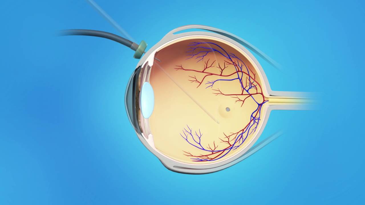
The most common surgical procedure for macular hole is a vitrectomy, which involves removing the gel-like substance in the center of the eye (the vitreous) and replacing it with a gas bubble. The bubble helps to push the retina against the back of the eye, which can help the hole to close and heal. The patient will need to maintain a specific head position for several days or weeks after the surgery to keep the gas bubble in the correct position.
After the surgery, the patient will need to use eye drops to prevent infection and reduce inflammation. They may also need to wear an eye patch for a few days to protect the eye while it heals. Vision may be blurry for several weeks after the surgery, but it should gradually improve over time.
In some cases, macular hole surgery may not be successful

